
About VITREORETINAL
The vitreoretinal centre in Mehta Eye Clinic is well equipped with state-of-art imaging and diagnostic facilities which deals with the management of diabetic retinopathy, ARMD, retinal detachment, trauma, infections and other retinal disorders.
We aspire to provide highest quality medical and surgical care for our patients with retinal diseases in a compassionate and efficient environment. Retinal related surgery is an extremely specialized and skilled area of practise. At Mehta Eye Clinic we have a dedicated team of specialists handling vitreoretinal disorders. Our doctors are well trained with surgical and diagnostics skills for treating such specific retinal problems.
Common Diagnostic Tests at Mehta Eye Clinic
Lasers
Indirect Ophthalmoscopic Laser Delivery System (LIO): This laser delivery system is mainly used for peripheral retinal lesions:
Common Retinal Diseases
1. Diabetes and Eye Problem
Diabetes mellitus is emerging as a major public health problem in India. It is a multisystem disorder which affects the heart, kidneys, peripheral nerves and the eyes. In the eyes, High levels of blood sugar can lead to early onset of Cataract and or can lead to a condition known as Diabetic Retinopathy.
2. Diabetic Retinopathy
Diabetic retinopathy is an eye problem that can be caused by either type 1 or type 2 diabetes mellitus. Retinopathy occurs when diabetes damages the tiny blood vessels in the retina which may leak fluid and blood. It’s a sight threatening condition, however if diagnosed and treated, vision loss can be prevented.
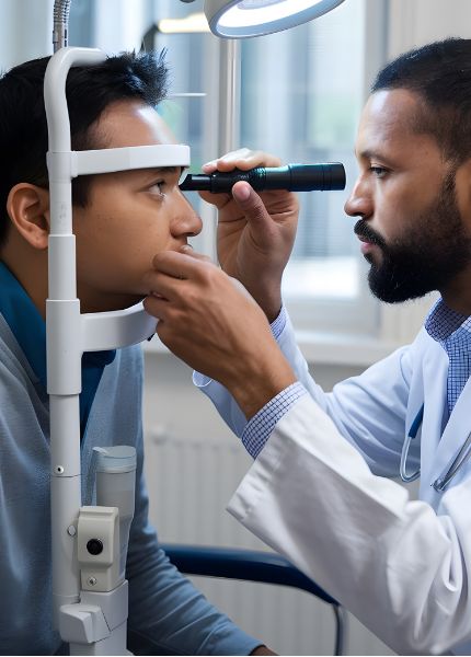
Firstly, examination of the retina of the eye is done using indirect ophthalmoscopy after dilating the pupils. If diabetic retinopathy is found, specialized test:
These Tests Will Help Us in Deciding the Stage & Treatment of Diabetic Retinopathy.
3. Vitrectomy
In the event of the patient presenting with very advanced diabetic retinopathy, a microsurgical procedure known as vitrectomy is recommended. Blood-filled vitreous gel of the eye is replaced with a clear solution to aid in restoring vision. Sometimes the retina may also be detached. Vitrectomy surgery is then performed to reattach the retina.
Sutureless Vitrectomy: Points to Remember
4. Age - Related MacularDegeneration (ARMD)
Age-related macular degeneration (ARMD) is the most common cause of irreversible vision loss in people over age of 60 years. Cells in the macula degenerate (the central, and most sensitive part of the retina at the back of the eye), that is, they become damaged and die. Damage to the macula affects your central vision which is needed for reading, writing, driving, recognizing people’s faces and doing other fine tasks.
There is NO TREATMENT for dry AMD, although high dose multivitamin combination has been shown to decrease the risk of visual loss.
There are a few treatment options for wet AMD although the best outcomes occur when this disease is detected early.
Not all patients may benefit from these, and treatment may not prevent further vision loss.
5. Retinal Detechment
The Retina:
The retina is a nerve layer at the back of the eye that senses light and sends images to the brain.
Meaning of Retinal Detachment:
The retina is the light-sensitive layer of tissue that lines the inside of the eye and sends visual messages through the optic nerve to the brain. When the retina detaches, it is lifted or pulled from its normal position. If not promptly treated, retinal detachment can cause permanent vision loss.
Retinal tears without detachment – Laser photocoagulation or cryotherapy is performed around the tear to seal the retina to the back wall of the eye.
Retinal tears with retinal detachment – Requires surgery as soon as possible to put the retina back in its proper position. The longer the retina stays detached, the less the visual improvement after surgery. Scleral buckle surgery – A flexible silicone band is placed around the eye to counter the force pulling on the retina after removing the fluid under the detached retina.
Retina Clinic FAQs
Are retinal injections painful?
Retinal injections are generally well tolerated. The eye is numbed with anesthetic drops before the procedure, which helps minimize discomfort. Most patients feel pressure rather than pain, and the procedure is completed quickly.
Can diabetic retinopathy be reversed?
Diabetic retinopathy cannot usually be reversed, but it can be effectively managed. Early detection and timely treatment help slow progression and preserve vision. Regular eye examinations are essential for people with diabetes.
Can retinal damage be repaired?
Some retinal conditions can be treated or stabilized, depending on the extent and cause of the damage. While not all retinal damage can be fully reversed, appropriate treatment can help protect remaining vision and prevent further deterioration.